Weldix® Suture Anchor System
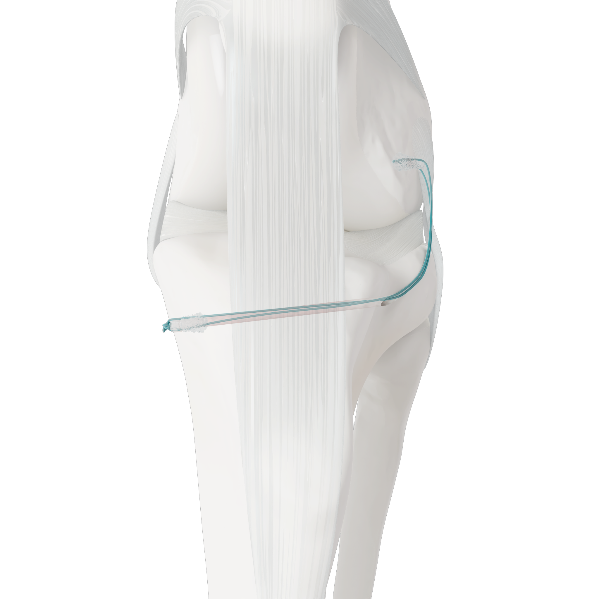
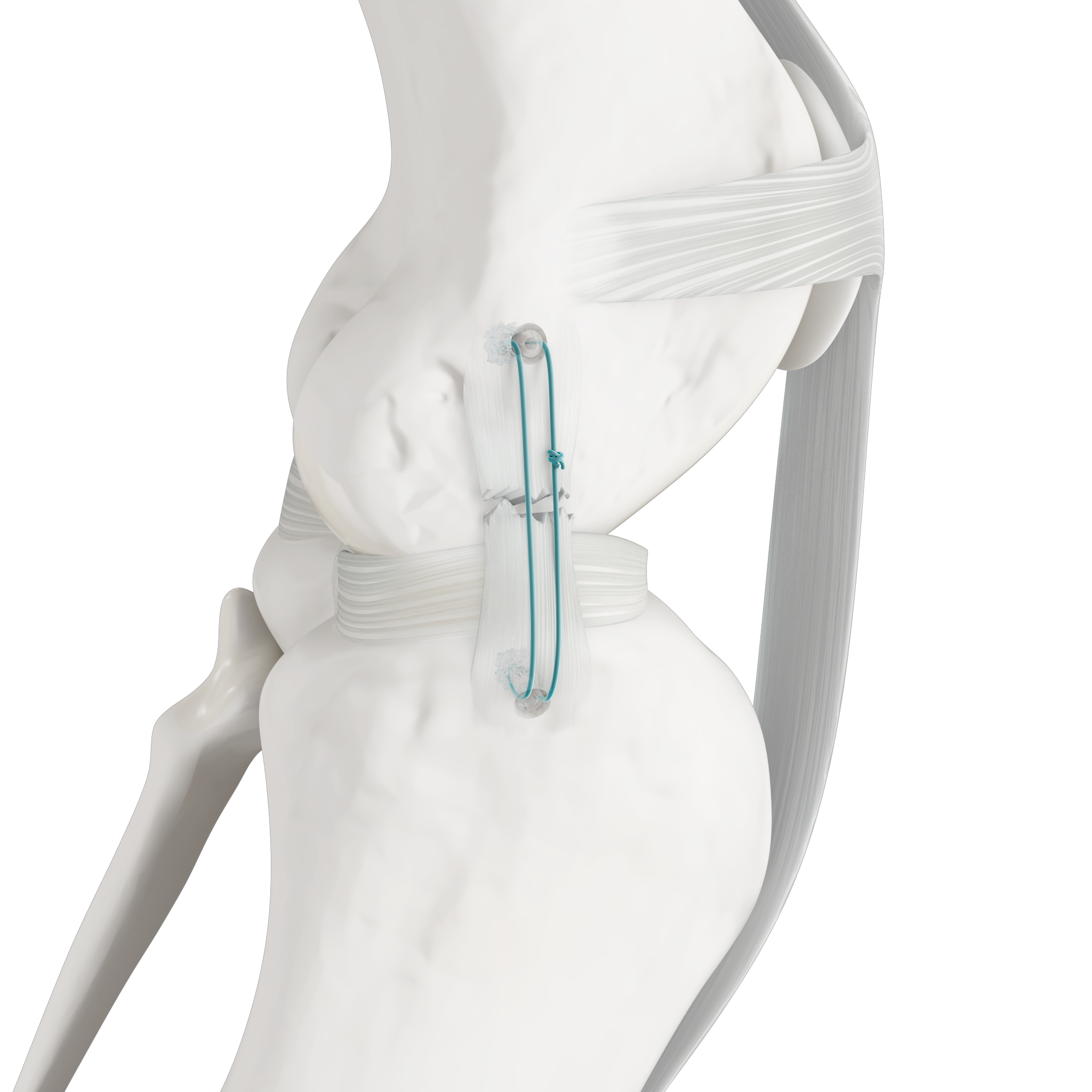
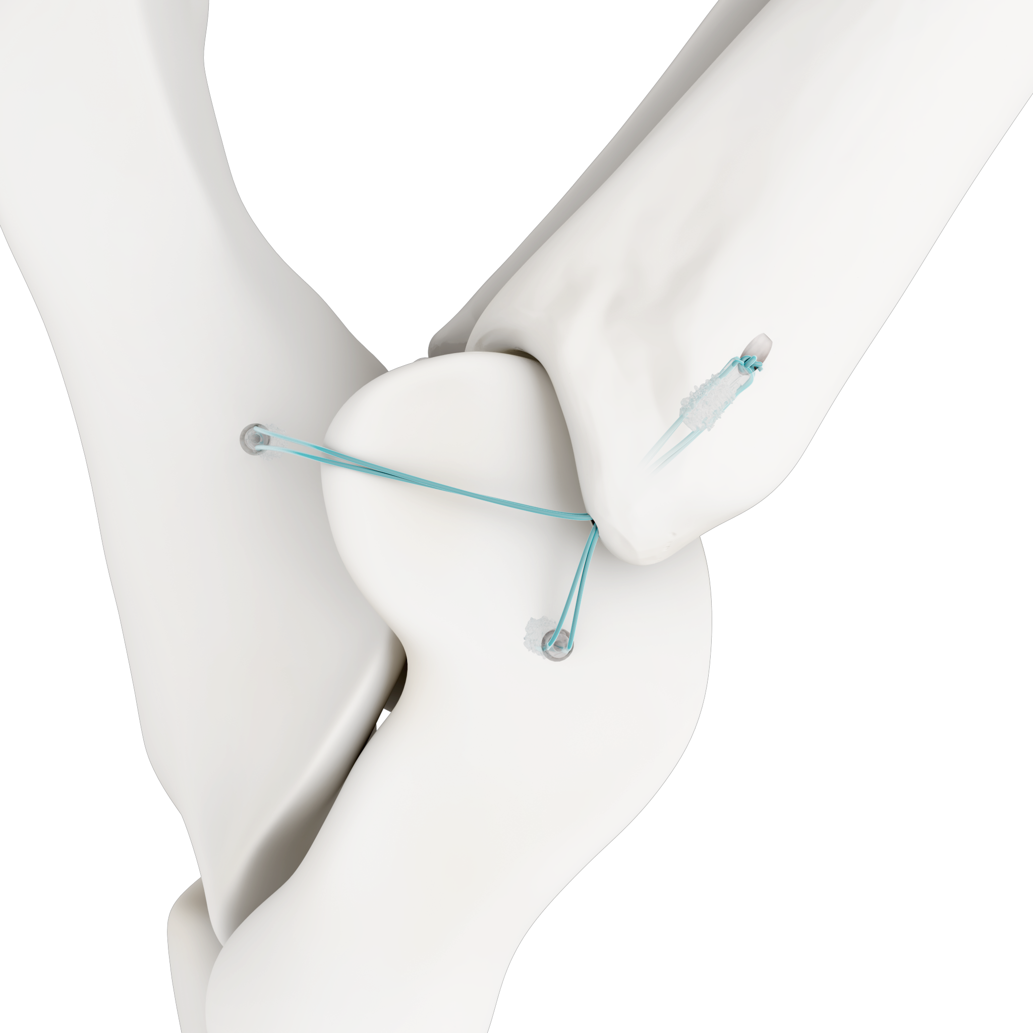
Resorbable - Reliable - Revolutionary

Excellent Anchorage 

Fully Resorbable 

Small Footprint 

Fast and Easy to use

Wide Range of Indications

The Weldix® Anchors can be used for several procedures in small animals such as cats and dogs, including:

  • Biceps tendon repair
  • Tarsal collateral ligament repair
  • Achilles tendon repair
  • CCL extracapsular repair
  • Knee collateral ligament repair
  • Elbow / Shoulder luxation repair
  • Hip luxation (capsule repair, ligamentum teres femoris repair)

Weight Chart:

Suture Compatibility

The Weldix® Anchors features one or two transverse apical “click-in” grooves for positioning the suture before implantation. The Weldix® Anchors can be used with the following sutures:

Weldix System Instruments

Ultrasonic Generator

The heart of the Insertion Technology is the BoneWelder Vet Ultrasonic System. The  System comprises a Handpiece that transmits the ultrasonic vibrations to the implants. The ultrasonic vibration is activated by a push-button directly on the handpiece.

BOS Drill

The Drill holes can be precisly  drilled with the light, ergonomic, battery powered BOS Drill

Weldix® Instrument Tray

Less Trauma bei Higher Strength

The Weldix® Suture Anchor requires only half the pilot hole size compared to standard anchors of similar pull-out strength.

Fast and Easy to use

The insertion of the anchor takes only a few seconds

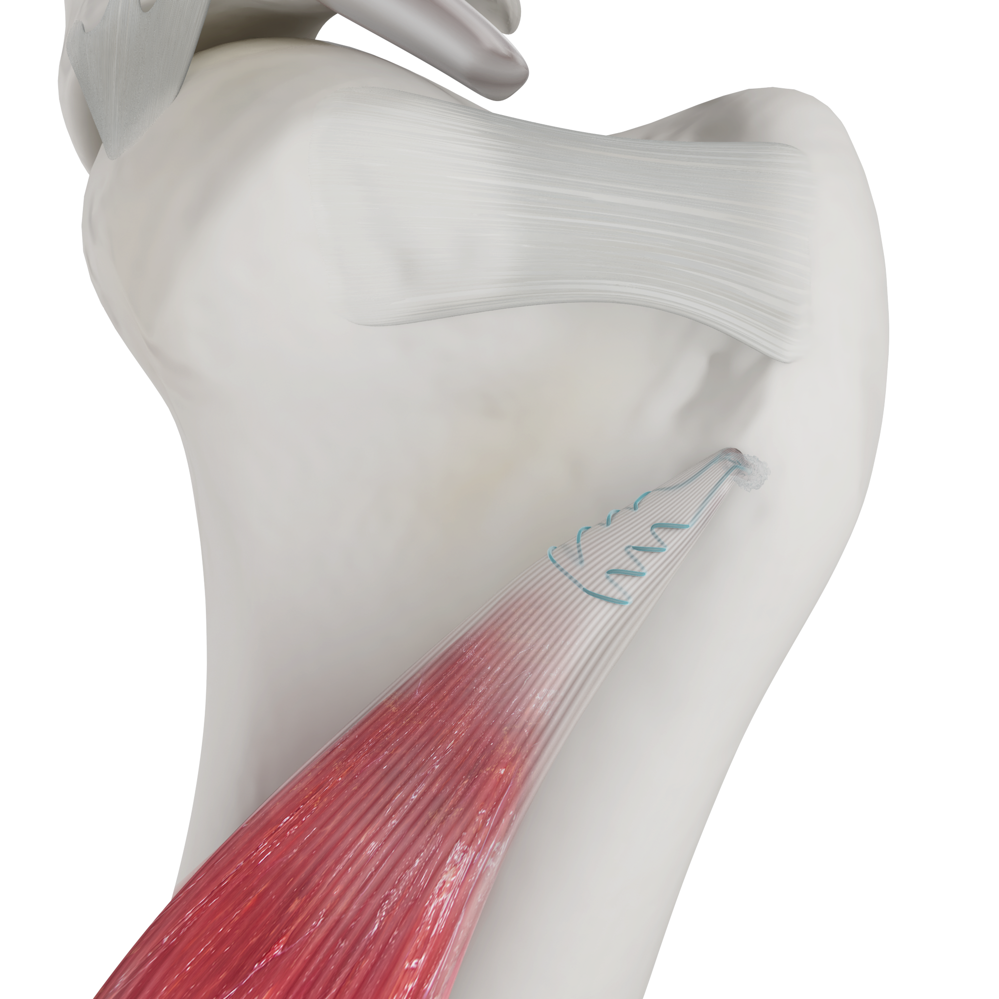
Ultrasonic energy is employed to liquefy the Weldix® Suture Anchor at the interface with bone tissue.
The liquid polymer flows into the marrow space of the surrounding cancellous bone where it is immediately quenched and provides strong anchorage of the implant.
The thermal impact on the bone is minimal and does not disturb bone healing and osseointegration.

Implantation Process

Drilling

A pilot hole is drilled into the bone to the depth of the integrated stop. The drill bit is equipped with a countersink to help position the anchor during implantation.

Mounting

The Weldix® 2.3mm Suture Anchor is placed on the tip of the sonotrode. Its precise geometry ensures a secure fit. The suture is clamped into the clip at the tip of the Weldix® 2.3mm Anchor and the anchor is positioned above the pre-drilled hole.

Implantation

Ultrasonic energy is applied, and the outer portion of the Weldix® 2.3mm Suture Anchor polymer liquefies. The polymer infiltrates the surrounding bone and immediately solidifies, locking both the implant and suture securely in place. Immediately after implantation, the suture is loadbearing.

Download Material

Publications

Koch, L.; Bockstahler, B.; Tichy, A.; Peham, C.; Schnabl-Feichter, E. Comparison of Extracapsular Stabilization Techniques Using an Ultrasonically Implanted Absorbable Bone Anchor (Weldix) after Cranial Cruciate Ligament Rupture in Cats—An In Vitro Study. Animals 2021, 11, 1695.

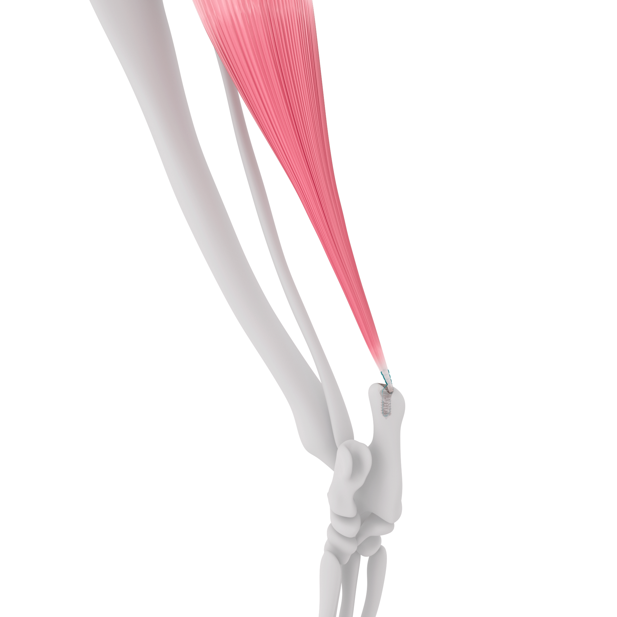
Schuenemann R, Strauss S. Biceps tenodesis with a bioabsorbable bone anchor using BoneWelding technology: Results in six clinical cases (5 dogs). Vet Surg. 2025 May;54(4):799-806. 

Trusted by Experienced Surgeons

Dr. Riccarda Schuenemann

Small Animal Hospital Sattledt, Austria

“The Weldix® 2.3mm Anchor is a promising technology with advantages over other anchor systems in regions with limited bone stock or soft tissue coverage and under bony protruberances.“

Dr. Riccarda Schuenemann

Small Animal Hospital Sattledt, Austria

“The Weldix® 2.3mm Anchor is a promising technology with advantages over other anchor systems in regions with limited bone stock or soft tissue coverage and under bony protruberances.“

previous arrow
next arrow
Slider

Let's get in touch!