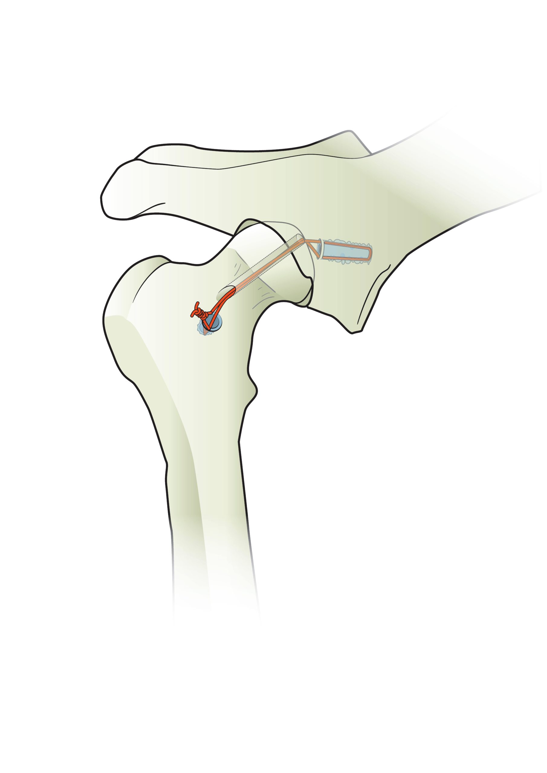
Weldix® Anchor - The revolutionary ultrasonic micro-suture anchor

Product overview

Providing excellent anchorage – even without cortical bone.
  • Fully resorbable
  • Outstanding strength
  • No micromotion
Dimensions

The Weldix® Anchor is 2.3mm in diameter and 7.2mm in length and requires a drill hole of 1.8mm in diameter and 7.6mm in depth.

Material

The implant is made of biocompatible and fully bioresorbable Polylactide (PLDLLA 70:30). The in vivo degradation of the Weldix® implant provides progressive loading during the healing process.

Degradation process

The Weldix® 2.3mm Anchor degrades due to the natural physiological process of hydrolysis, which results in a complete metabolization of the polymer into H2O and CO2 over 18 months.

Indications

The Weldix® 2.3mm Anchor can be used for several procedures in cats and dogs, including:

  • Hip luxation (capsule repair, ligamentum teres femoris repair)
  • Tarsal collateral ligament repair
  • Achilles tendon repair
  • CCL extracapsular repair
  • Knee collateral ligament repair
  • Elbow / Shoulder luxation repair

Weldix® 2.3mm Anchor Pullout Strength

The Weldix® 2.3mm Anchor shows superior pullout strength compared to conventional anchors

All Suture Anchor: Anchor made from suture only, forming a knot in the bone while tightening

Mechanical Suture Anchor: Titanium anchor having mechanical retention structures like barbs or threads to create a hold in bone

Sutures

The Weldix® 2.3mm Anchor can be used in combination with the following sutures:

Sutures are not included in the package. 

Suture clip function

The Weldix® 2.3mm Anchor features two transverse apical “click-in” grooves for positioning the suture before implantation.

Ultrasonic Generator

Weldix® Anchors must be implanted with the BoneWelder® Vet ultrasonic generator.

Weldix® Twist Drill

The twist drill (with or without drill stop) is sized specifically for the Weldix® 2.3mm Anchor. It helps ensure safe and easy handling of the implants and correct implantation.

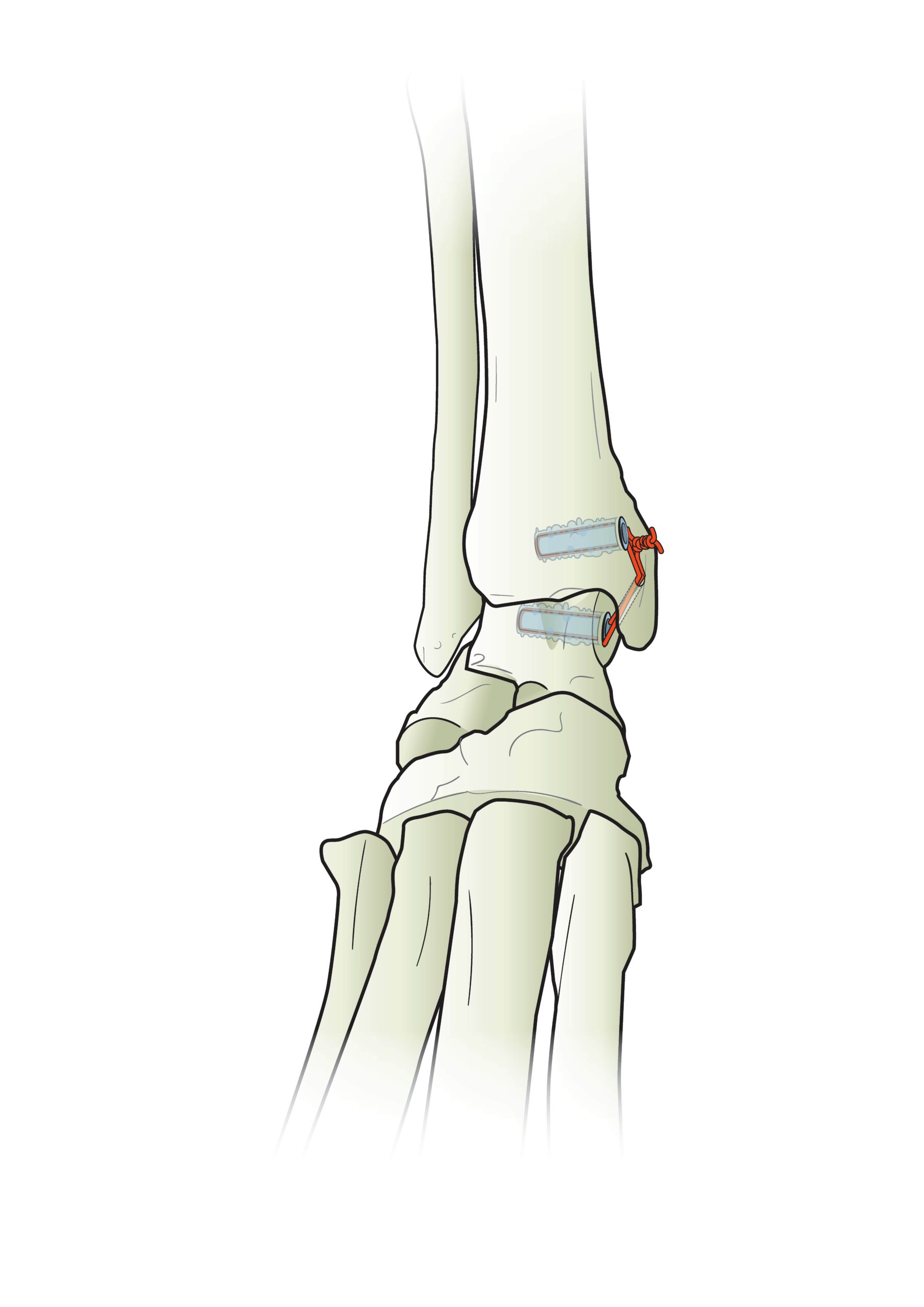
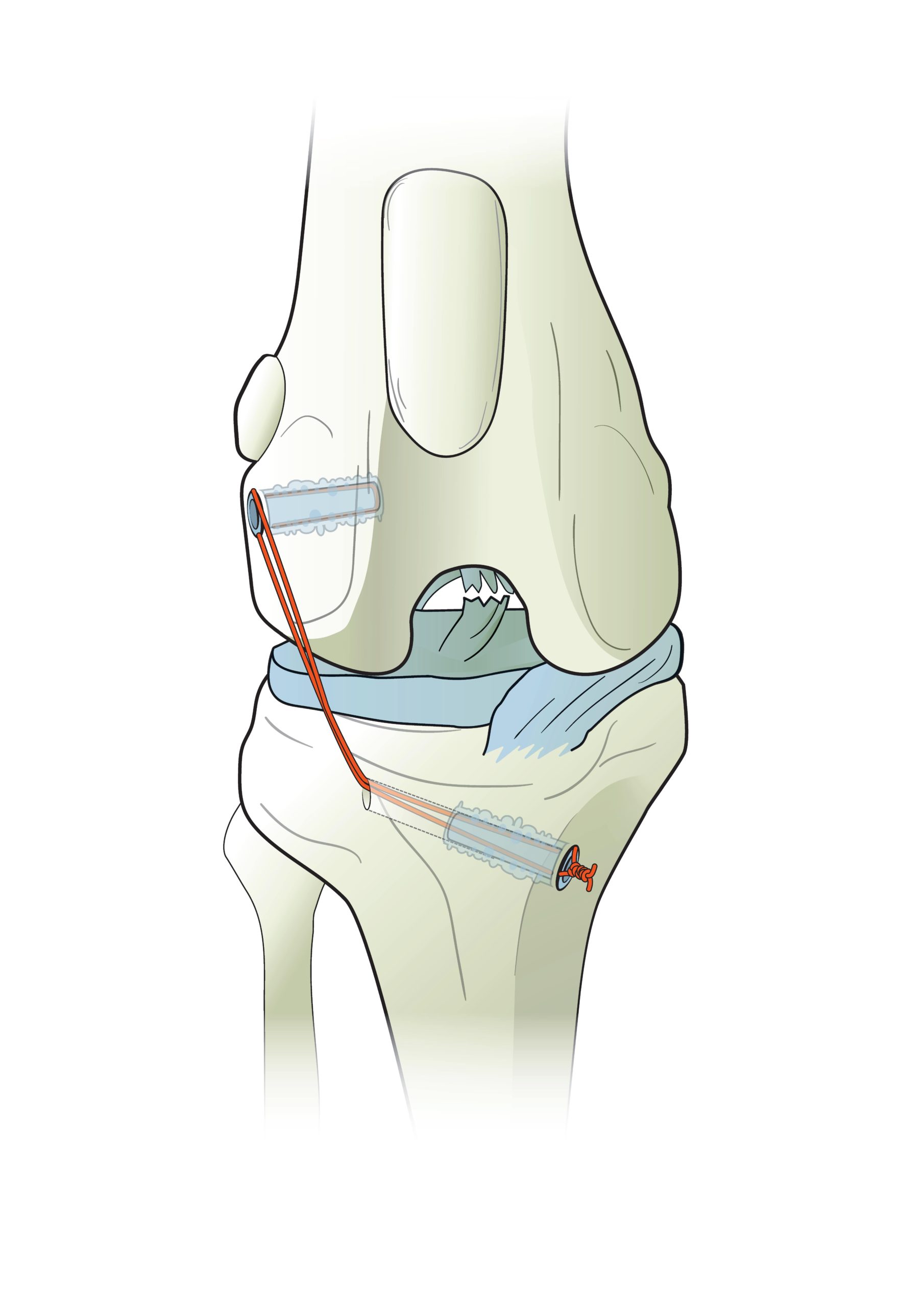
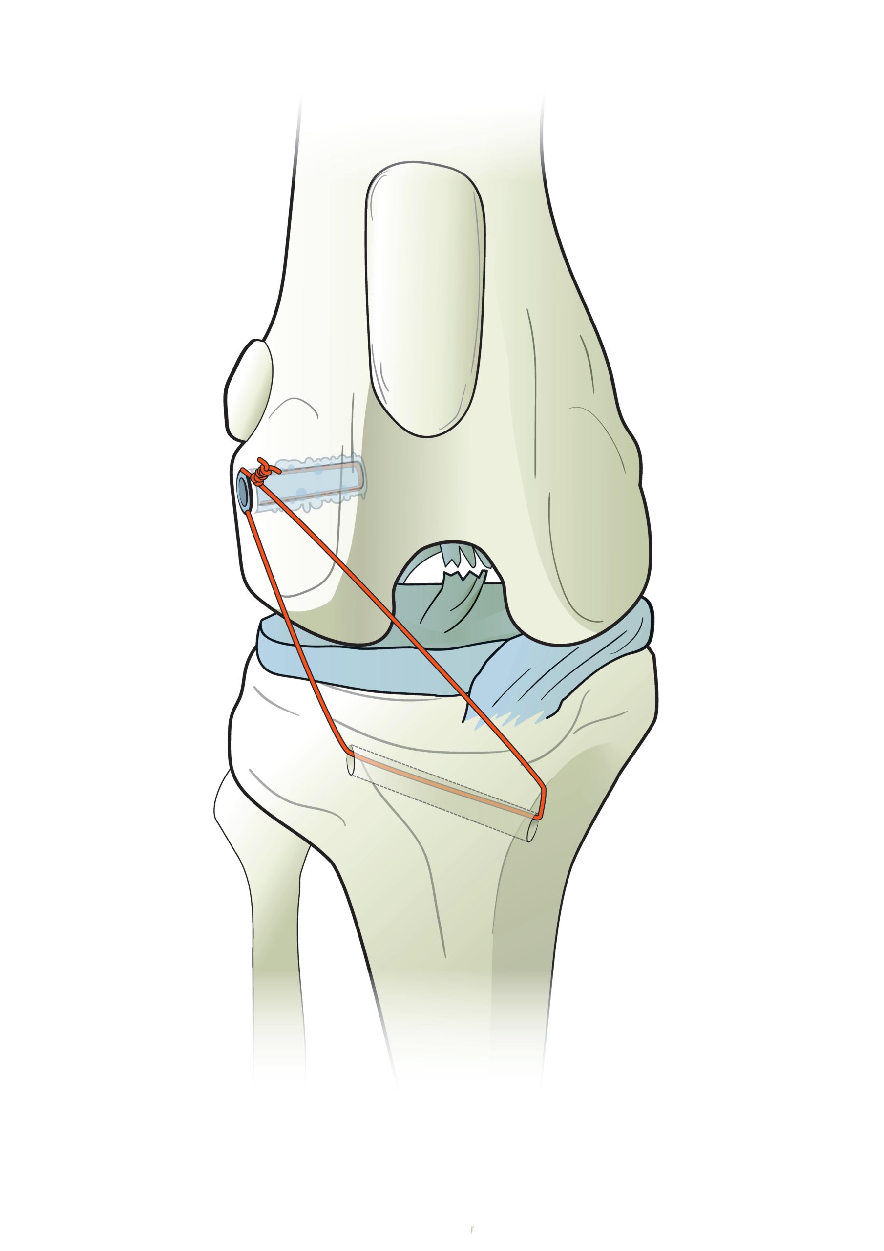
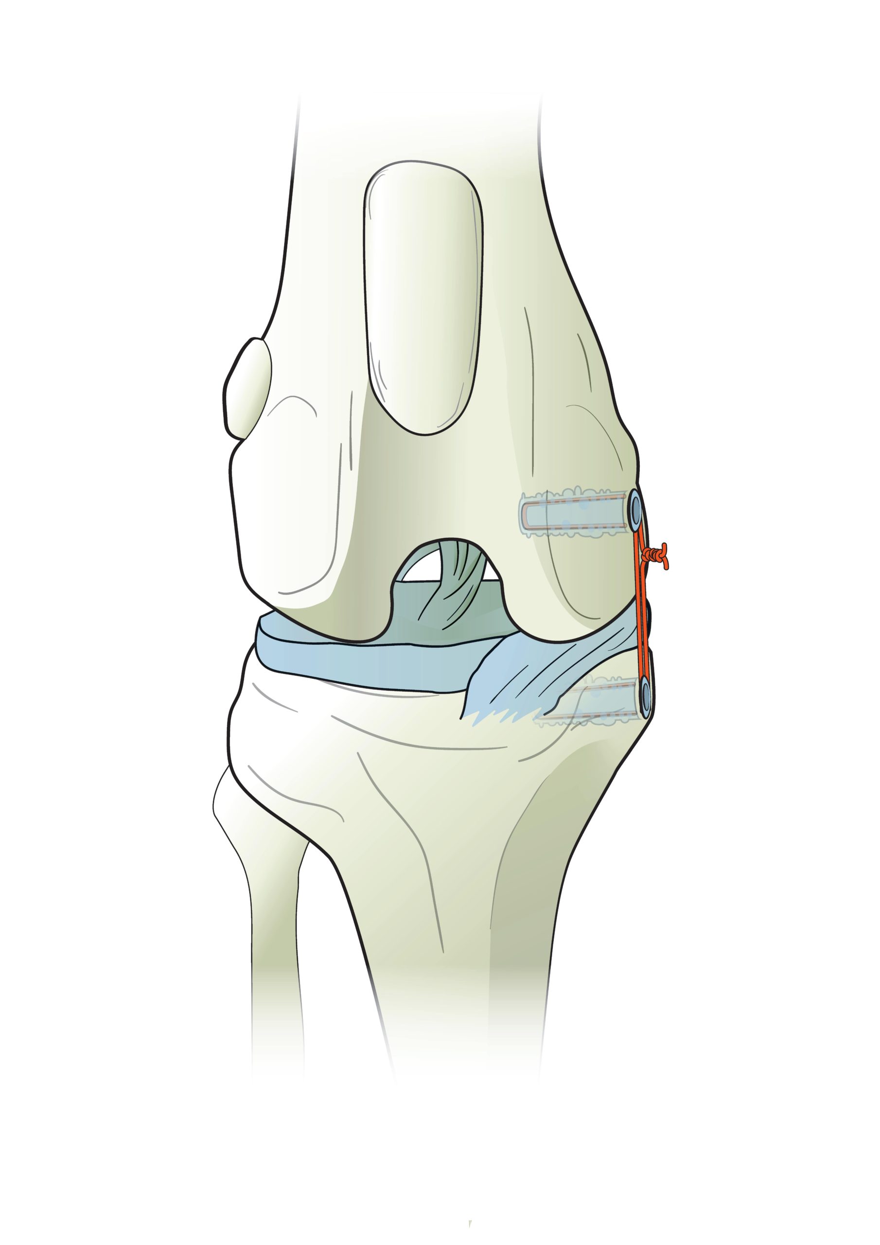
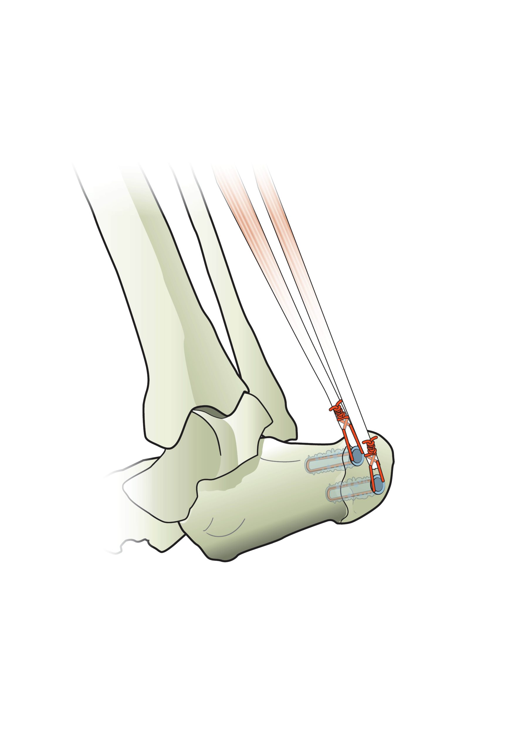
Animated Implantation Process for the

Weldix® Suture Anchor

Implantation Process

Drilling

A pilot hole is drilled into the bone to the depth of the integrated stop. The drill bit is equipped with a countersink to help position the anchor during implantation. 

Mounting

The Weldix® 2.3mm Suture Anchor is placed on the tip of the sonotrode. Its precise geometry ensures a secure fit. The suture is clamped into the clip at the tip of the Weldix® 2.3mm Anchor and the anchor is positioned above the pre-drilled hole.

Implantation

Ultrasonic energy is applied, and the outer portion of the Weldix® 2.3mm Suture Anchor polymer liquefies. The polymer infiltrates the surrounding bone and immediately solidifies, locking both the implant and suture securely in place. Immediately after implantation, the suture is loadbearing.

Weldix® Anchor Download Material

Trusted by Experienced Surgeons

Dr. Riccarda Schuenemann

Small Animal Hospital Sattledt, Austria

“The Weldix® 2.3mm Anchor is a promising technology with advantages over other anchor systems in regions with limited bone stock or soft tissue coverage and under bony protruberances.“

Dr. Riccarda Schuenemann

Small Animal Hospital Sattledt, Austria

“The Weldix® 2.3mm Anchor is a promising technology with advantages over other anchor systems in regions with limited bone stock or soft tissue coverage and under bony protruberances.“

previous arrow
next arrow
Slider

Let's get in touch!